
Endourology Survey on Radiation Exposure Reveals A Need for Clear Guidelines for Post Ureteroscopy Imaging
Ohad Kott, MD1, Osama Al-Alao, MD1, Jorge Pereira, MD2, Christopher Tucci, RN1, Gyan Pareek, MD1
1Minimally Invasive Urology Institute, The Miriam Hospital; Department of Urology, The Warren Alpert Medical School of Brown University, Providence, RI, 2Columbia University Division of Urology, Mount Sinai Medical Center, Miami Beach, FL
BACKGROUND: Nephrolithiasis patients undergo repeated imaging studies throughout their lives, exposing them to large doses of radiation, potentially leading to secondary malignancies. Studies have found that effective dose of radiation exceeded the recommended levels in up to 20% of nephrolithiasis patients throughout the evaluation and follow up period. Currently, guidelines only suggest recommendations regarding postoperative imaging following ureteroscopic lithotripsy (URSL) and strategies to minimize radiation exposure. There are no recommendations on the frequency and modality of imaging utilized in stone formers. The latter varies and depends on practitioner discretion. As such, we sought to elucidate the common imaging practices following URSL and current knowledge of radiation exposure among endourologists.
METHODS: A 15-item web-based survey was conducted among active members of the Endourological Society. The survey evaluated knowledge and perception of best practice and patient radiation exposure in post URSL imaging. The survey also collected clinical volume, training, experience and location of practice.
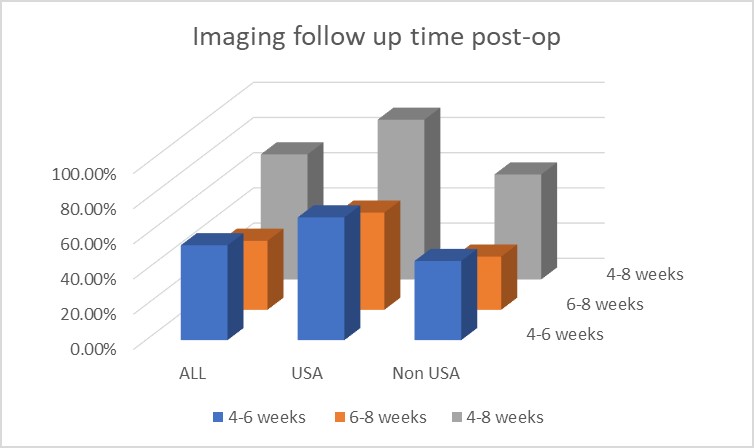
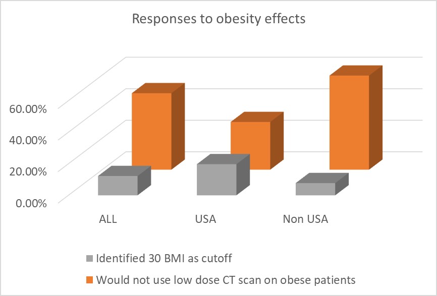
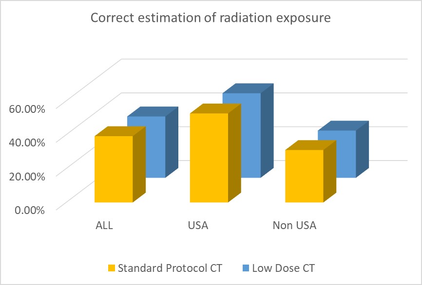
RESULTS: 204 endourologists completed the survey with a mean of 13.29 years in practice (IQR 6.75, 20 years). Routine postoperative follow up imaging is regularly performed by 91.7% of respondents using the following modalities: Ultrasound (US) - 76.6%, X-ray (KUB)- 44.7%, computerized tomography (CT) - 39.6%. 53.92% of respondents reported performing follow up imaging between 4-6 weeks, while 39.22% cite between 6-8 weeks and 71.08% reported between 4-8 weeks. 78.43% of respondents consider the imaging quality of low dose CT scan similar to standard dose renal stone protocol CT (SP CT). 48.53% of respondents would not use low dose CT scan on obese patients while only 12.25% correctly identified that streak effect is significant for BMI>30 Kg/m2. 39.22% of respondents estimated correctly the radiation exposure of SP CT scan while 36.27% of respondents estimated correctly the radiation exposure of a low dose CT scan.
CONCLUSIONS: Our data reveals that urologists worldwide need better education on radiation exposure from diagnostic studies. Additionally, there appears to be a knowledge deficit with the timing and utilization of low dose CT. This deficit appears larger outside than within USA. Current guidelines appear to be discretionary, not supported with enough evidence and do not take into account the utilization of low-dose CT. Clear evidence-based guidelines may help alleviate these knowledge gaps and undue radiation exposure to nephrolithiasis patients.
Table:
| ALL | USA | Non-USA | ||
| Completed the survey | 204 | 76 | 129 | |
| Years in practice (IQR) | 13.29 (6.75, 20) | 11.52 (6.5, 15) | 14.29 (6.75, 20.25) | |
| Perform routine follow up imaging | 91.67% | 97.30% | 88.37% | |
| US | 76.60% | 91.90% | 67.20% | |
| KUB | 44.70% | 45.90% | 44.30% | |
| CT | 39.60% | 50% | 32.80% | |
| How long after surgery do you perform imaging (in weeks)? | ||||
| 4-6 weeks | 53.92% | 69.74% | 44.96% | |
| 6-8 weeks | 39.22% | 55.26% | 30.23% | |
| 4-8 weeks | 71.08% | 90.79% | 59.69% | |
| Low dose CT imaging quality is similar to SP | 78.43% | 77.63% | 78.29% | |
| Would not use low dose CT scan on obese patients | 48.53% | 30.26% | 59.69% | |
| Identified 30 BMI as cutoff for streak effects | 12.25% | 19.74% | 7.75% | |
| Estimated correctly the radiation exposure in: | ||||
| Standard Protocol CT | 39.22% | 52.63% | 31.01% | |
| Low Dose CT | 36.27% | 50.00% | 27.91% | |

Back to 2019 Abstracts