Back to 2025 Abstracts
Renal pseudoaneurysm identification: a real-world sensitivity of pre-angiogram imaging
Anna Saltman, MD, MS
1,
Samuel Iofel, BA2, William Faust, MD
1.
1Lahey, Burlington, MA, USA,
2Tufts University School of Medicine, Boston, MA, USA.
Introduction:Renal pseudoaneurysm (rPSA) is a rare but life-threatening complication following percutaneous renal procedures, with an incidence of 0.36%-5%. Standard management involves digital subtraction angiography (DSA) with embolization. While DSA is the gold standard for diagnosis and treatment, many patients receive imaging before this intervention, with the sensitivity of CT angiogram (CTA) reported at 90.3%. This adds IV contrast load and may delay treatment. Our study evaluates the clinical utility and impact of pre-angiography imaging on postprocedural outcomes.
Methods:After IRB approval, we reviewed patients with suspected post-procedure rPSA from 2015-2024. Procedures included percutaneous nephrolithotripsy (PCNL), nephrostomy tube placement, renal biopsy, and cryoablation. rPSA suspicion was based on hematuria, hematocrit drop, or perinephric hematoma. Data on demographics, imaging type, pre/post-procedure hematocrit and creatinine, length of stay, and blood transfusions were collected. Statistical analysis included t-tests, ANOVA, and Fisher�s Exact test. Imaging sensitivity was compared using DSA as the reference.
Results:
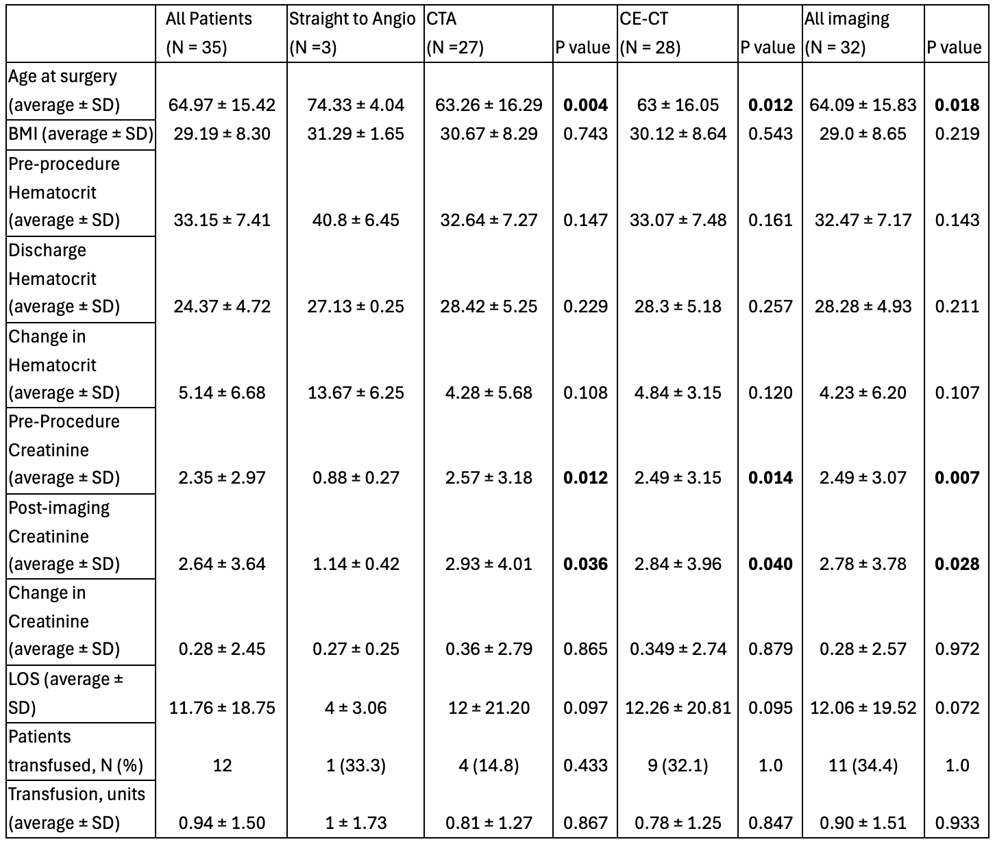
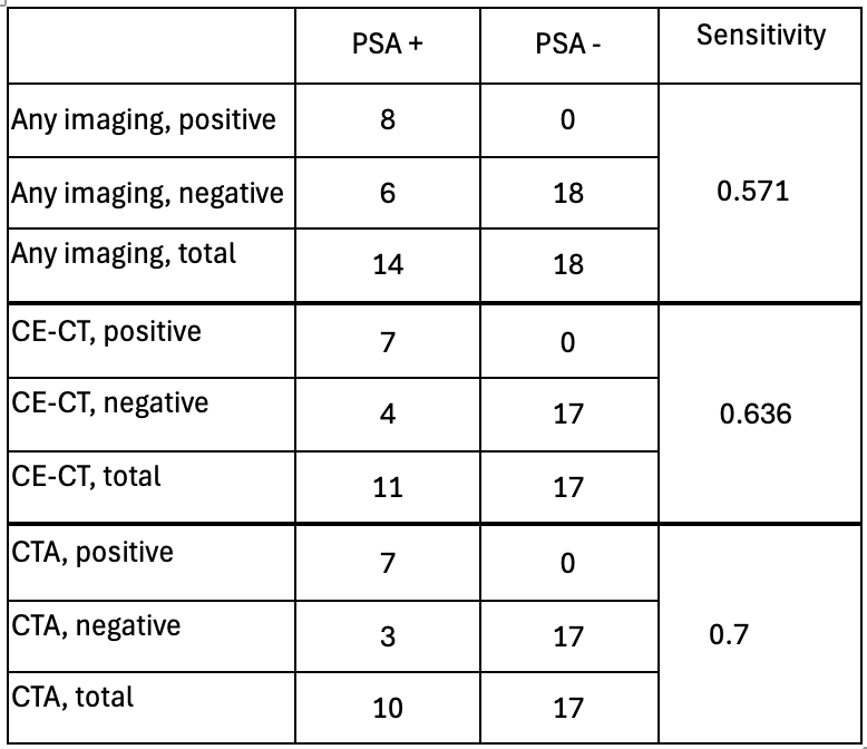
Among 38 rPSA patients, mean age was 64.97 years, and BMI was 29.19 kg/m�. Hematocrit dropped by 5.14% (p < 0.0001), and creatinine increased by +0.4 mg/dL (p = 0.25). Sensitivity of pre-DSA imaging varied: any preop imaging detected 57.1% of rPSA, CE-CT 63.6%, and CTA 70%. Patients undergoing direct DSA (N=3) were older (74.33 years) but had similar BMI and clinical outcomes.
Conclusion:CTA sensitivity was lower than reported in the literature, especially when accounting for non-contrast-enhanced CT. Despite limitations in sample size, our findings suggest that pre-DSA CT imaging may have limited benefit for patients with high clinical suspicion of rPSA.
Back to 2025 Abstracts


