|
Back to 2011 Program
Measurement of Spatial Distribution in Prostate Biopsy
Misop Han, Chunwoo Kim, Doyoung Chang, Hyungju Kim, Doru Petrisor, Dan Stoianovici
Johns Hopkins Medical Institutions, Baltimore, MD
Introduction: Prostate biopsy is typically performed freehand with transrectal ultrasound (TRUS) guidance. However, it is difficult to determine the accuracy of the spatial distribution of TRUS-guided biopsy.
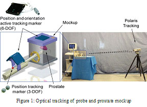
Materials & Methods: A simulation model was built to accurately measure in-vitro biopsy locations. This model consists of (1) a gelatin-based pelvic mockup with precisely defined geometry of a prostate and rectal cavity, (2) an optical tracking system (Polaris®, NDI, Ontario, Canada) to measure the relative locations, (3) TRUS probe, and (4) biopsy needle (Figure 1). Overall, this system measures the precise locations where biopsy cores are sampled. The measurement errors of the instrument are within 1 mm. After defining the gold standard using a 12-core sextant biopsy plan, we determined how closely an experienced urologist can perform a biopsy compared to the gold standard.
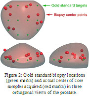
Results: Results from a simulated biopsy are shown in Figure 2. The simulated biopsy cores were often clustered and a large portion of the prostate gland was undersampled. The average error distance was 8.86 mm.
Conclusions: TRUS-guided prostate biopsies may not closely follow sextant biopsy distribution. An alternate targeting method may be needed for uniform sampling during TRUS-guided prostate biopsy.
 
Back to 2011 Program
|